
Experimental principle
As the DNA double-stranded molecule is continuously increased in temperature or treated with chemical denaturing agents, the two strands begin to separate (ie, melt). The first melting zone consists of bases with a lower melting temperature. GC bases are strongly bonded to AT base pairs, so regions with high GC content have higher melting temperatures. The factors that affect the melting temperature at the same time are the attractive forces between adjacent bases. The region where the melting temperature is low, usually at the end, is called the low temperature melting zone. If the end is partially open, the double helix is ​​bundled together by the unmelted part, and this area is called high temperature melting (Fig. 1).
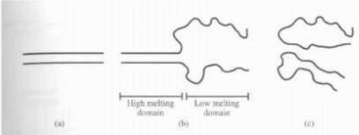
figure 1
If the temperature or denaturant concentration continues to rise, the two chains will be completely separated. The first point of denaturing gradient gel electrophoresis is that once the DNA double-stranded ends are melted, the electrophoresis speed in the gel will drop dramatically. The second basis is that if a region is first melted, and another chain with only one base difference will have a different melting temperature, the electrophoresis speed will also be significantly different. Therefore, the sample is added to a gel containing a denaturant gradient for electrophoresis to separate the two (Figures 2, 1 and 2).

figure 2
Finally, if a double strand is in its low temperature melting zone base mismatch (heteroduplex), and the difference is only in this case compared to another equivalent double strand, then the double strand containing the mismatched base will be low. Much of the denaturing concentration is melted. In fact, samples typically contain mutations, normal homoduplexes, and paired heteroduplexes, which are produced when PCR is amplified. The double strands containing mismatches (lanes 3 and 4 in Figure 2) can usually be separated from two homologous duplexes (lanes 1 and 2 in Figure 2). This separation makes the method highly sensitive. . In order to achieve the best separation of different molecules with only one base difference, it is necessary to first select the range of DNA to be studied and the concentration gradient of the denaturant during the electrophoresis sample. This can be empirically solved by the orthogonal denaturation gradient experiment shown in Figure 2. The denaturant gradient should be chosen in the portion where the slope of the curve is large, since most of the molecules are in a partially denatured state, which allows optimal separation of the different molecules falling into the low temperature melting zone.
Most analytical experiments now include the addition of a "GC clamp". It is a 30-50 base, GC-rich DNA attached to one end of the double strand to form an artificial high temperature melting zone. In this way, the rest of the fragment is in the low temperature melting zone so that it can be analyzed.
Experimental Materials
1. Experimental instrument: one electrophoresis temperature control tank, electrophoresis support, programmable electrophoresis apparatus, gradient mixer, buffer siphon pump, gel imager;
2. Experimental reagents:
(1) 40% of gel monomer (acrylamide/methylene bisacrylamide: 37.5/1): Weigh 38.93g of acrylamide and 1.07g of methylidene bisacrylamide, add to deionized water to make up to 100ml The 0.45 μm filter was filtered and placed in a brown bottle and stored at 4 ° C.
(2) 100% denaturing agent: Take 42 ml of deionized formamide and 42 g of urea, dilute to 100 ml with deionized water, filter with a 0.45 μm filter, and store in a brown bottle at 4 ° C.
(3) 50 × TAE buffer (Tris 2mol / L, glacial acetic acid 1mol / L, EDTA 50mmol / L, pH 8.0);
(4) 10% ammonium persulfate solution (W / V, in order to ensure a good triggering effect, it is recommended to use now);
(5) TEMED (analytical grade).
(6) Formulation of gradient denatured gel (denatility gel with a concentration of 8%)
Component | Denaturant concentration 60% | Denaturant concentration 50% | Denaturant concentration 25% | Denaturant concentration 0% |
Gel monomer | 2.8ml | 2.8ml | 2.8ml | 0.8ml |
100% denaturant | 8.4ml | 7ml | 3.5ml | 0 |
50X TAE | 0.28ml | 0.28ml | 0.28 | 0.08ml |
Double distilled water | 2.52ml | 3.92ml | 7.42ml | 3.12ml |
10% ammonium persulfate | 60ul | 60ul | 60ul | 25ul |
TEMED | 8ul | 60ul | 60ul | 5ul |
total capacity | 14ml | 14ml | 14ml | 14ml |
(7) Formulation of gradient denatured gel (denatility gel with a concentration of 6%)
Component | Denaturant concentration 60% | Denaturant concentration 50% | Denaturant concentration 25% | Denaturant concentration 0% |
Gel monomer | 2.1ml | 2.1ml | 2.1ml | 0.6ml |
100% denaturant | 8.4ml | 7ml | 3.5ml | 0 |
50X TAE | 0.28ml | 0.28ml | 0.28 | 0.08ml |
Double distilled water | 3.22ml | 4.62ml | 8.12ml | 3.32ml |
10% ammonium persulfate | 60ul | 60ul | 60ul | 25ul |
TEMED | 8ul | 60ul | 60ul | 5ul |
total capacity | 14ml | 14ml | 14ml | 14ml |

experiment procedure
1. Align the two pieces of glass into the electrophoresis holder (the notched glass is facing up and on the inside), tighten the set screw and clamp the clip. Inject deionized water to detect if water is leaking and then pour out the ionized water.
2. Prepare a gel solution with a high denaturant concentration and a gel solution with a low denaturant concentration. Add a proper amount of 10% ammonium persulfate solution and TEMED as a polymerization initiator and catalyst before mixing and mix well. Close the two valves of the gradient mixer, add the high concentration denaturing gel solution to the end of the gradient mixer near the outlet, add the low concentration denaturing gel solution to the other end, and turn on the magnetic stirrer.
3. First open the outlet end of the gradient mixer. When the flow is stable and no bubbles, insert the needle into the middle of the two glass gaps, and then open the middle of the gradient mixer to start the filling.
4. Stop the gelation when the gel is poured 1.5cm from the upper end, add 1mL deionized water to seal the rubber surface, and after gel polymerization, prepare 0% gel without denaturing agent, and fill the glue with pipette. Insert the comb until the gel is completely polymerized.
5. Pull out the comb, place the electrophoresis scaffold into the thermostatic electrophoresis tank, mix each PCR product with the Loading buffer, then aspirate the sample with a micro-sampler and add it to each well (the DNA content of each well is 500). -800 ng, the PCR product was compared with Marker in agarose electrophoresis to calculate the approximate content, so that the DNA content of each sample of the gradient densified gel was as uniform as possible). After everything is ready, start to start electrophoresis, the voltage is set to 60V in the first stage, time 1h, second stage 100V, time 16h, electrophoresis automatic timed segmentation programming.
6. After the electrophoresis is finished, take out the electrophoresis frame and take out the glass, peel off the glass on one side, and then remove the gel after staining with EB or SYBR Green for 30 minutes, and take a photo in a gel imager.
Precautions
1. Extract DNA during the experiment, and contact with more volatile organic reagents and toxic reagents during DGGE operation. Pay attention to protection during operation.
2. The gel concentration is selected according to the length of the PCR product fragment, generally 8% of the glue is selected for 150-400 bp, and 6% of the glue is used for 400 bp or more.
3. The concentration range of the denaturant is appropriately adjusted according to the literature and specific samples, so that the final strip is located in the middle of the denatured rubber.
4, the specific amount of initiator and catalyst can be adjusted according to the temperature and the experimenter's requirements for the polymerization rate and other specific experimental conditions, the reference amount is listed in the document table.
5, the glass plate used for the glue must be cleaned to avoid the impurities caused by air bubbles and other factors affecting the integrity of the gel, before adding the gel solution, it is necessary to check whether the glass plate leaks.
6. When taking out the comb, be careful not to damage the sample hole. You can first soak the gel in the running buffer and then remove it.
7. Insert 1×TAE buffer 21L into the electrophoresis tank in advance, set the temperature to 60 °C, insert the electrode and buffer circulation tube, and preheat the electrophoresis buffer.

Remarks: Weizheng Biotechnology Co., Ltd. is committed to providing high quality virus packaging services for researchers. Services include: scientific and clinical grade adenovirus, lentivirus, adeno-associated virus (AAV) packaging, plasmid vector construction, TALEN gene Knockout, gene mutation, etc. So far, the company has a human-source stock database library (18,000), an adenovirus stock library (12,000), and an adeno-associated virus (AAV) stock library. The company also has a wealth of customized services. Sincerely welcome you to consult and purchase!
Antimicrobial Hemodialysis Catheter
Hemodialysis Catheter,dialysis Catheter,powertrialysis dialysis catheter,peritoneal dialysis catheter,dialysis catheter kit
Anesthesia Medical Co., Ltd. , https://www.trustfulmedical.com