Fura-2 QBT Kit - New Phase Fura-2 Calcium Flow Detection Experiment
Fura-2 dyes have long been recognized as an important tool for detecting calcium mobilization in experiments such as cell imaging, GPCR- mediated intracellular calcium flux, and ion channel activation. This ratiometric method detects dyes by calculating the ratio of fluorescence intensities between the bound and unbound states, helping to correct for errors due to dye addition or cell plating. However, conventional Fura-2 dyes must be buffer cleaned, thereby increasing inter-well variability for each experiment and requiring more time and increased operational complexity.
 Molecular Devices® introduced Fura-2 QBT ™ kit is based on calcium flux signal annihilation patented technology, containing calcium indicator Fura-2 ratio detection method provides a homogeneous system and reduce the difference in the experimental cell basis, so that Flux is increased indirectly by removing the cell wash step prior to detection. In addition , the Fura-2 QBT. Calcium Flow Kit is excited at the UV spectrum to maximize the interference of the compound's fluorescent signal at 488 nm .
Features:
Get a larger signal window based on signal annihilation techniques
No-clean and ratiometric signal detection minimizes experimental variability
The no-clean step saves time and experiment costs
Experimental principle
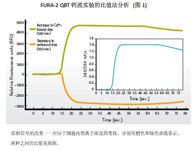
Fura-2 AM is a calcium-sensitive dye that is excited at 340 nm and 380 nm with an emission wavelength of 510 nm . When the dye binds to calcium ions, its absorption wavelength shifts from 380 nm to 340 nm . Figure 1 shows the increase in calcium ion binding 340 / 510nm signal is transmitted after the Fura-2 (green) and the signal (orange) 380/510 nm emission decreases. The ratio of the two transmitted signals (inset) is calculated to detect the signal effect after fitting the maximum value of the dose-effect curve minus the minimum.
Fura-2 QBT. Calcium flow experiment preparation
The small-scale Fura-2 QBT. Calcium Flow Kit ( PN #R8197 ) was used in this experiment . Resuspend the dye in 20 mM
HEPES, Hank 's balanced salt solution (HBSS). Probenecid ( PBX ) is added as needed to inhibit the dye from being transferred out of the cell. The other CHO M1 cells or HeLa cells were seeded overnight in a black-walled 384- well plate and removed from the incubator during the test. 25 μ L buffer containing Fura-2 QBT dyes or other dyes added to each well, and the wells contained 25 μ L required buffer or medium. Cell recording plates loaded with Fura-2 QBT. Calcium Flow Reagent ( PN #R8197 ) or BD Ratio Calcium Flow Reagent ( #644243 ) were incubated for one hour at 37 ° C , 5% CO 2 . For ease of comparison, the traditional Fura-2 cleaning method is used.

According to this conventional cleaning method Fura-2, the medium was removed from each well prior to the addition of 50 μ L dye 1X buffer, and then to other cells as recording kit version incubated under the same conditions and time. All recording plates were removed from the incubator and allowed to cool to room temperature for 10 minutes before recording was started on the FlexStation® 3 Microplate Reader or FLIPR® Tetra System . A recording plate using the conventional Fura-2 cleaning method needs to be washed 3 times with HBSS test buffer to remove excess dye before starting recording .
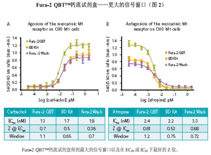
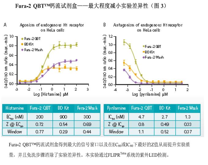
Ratio Calcium Flow Assay on FlexStation 3 and FLIPR Tetra Systems A 5- fold concentration of the corresponding ligand was dissolved in HBSS buffer containing 20 mM HEPES in a 384- well polypropylene compound plate . Agonists were added during the FlexStation 3 (Figure 2 ) or FLIPRTetra system (Figure 3 ) and the instrument parameters were optimized to fit the ratio test. The excitation wavelength of the dye is 340 nm and 380 nm . The emission signal was detected at 510 nm and the relative fluorescence units ( RFU ) at two excitation wavelengths in each well were recorded for approximately 90 seconds, including before and after loading. The calculated output at each sampling time point in the experiment is the ratio of the 340 nm to 380 nm wavelength signals. The calculation of the maximum value minus the minimum value is derived from the ratio signal curve. The data required for the dose-response curve and EC50/IC50 concentration calculations are exported from ScreenWorks® or SoftMax® Pro software and then further fitted and calculated using GraphPad Prism . The calculation of the .Z value was performed according to the method published by Zhang, et .
result


in conclusion
Compared to traditional Fura-2 experiments, the Fura-2 QBT. Calcium Flow Kit provides a homogeneous experimental system that eliminates the need to rinse cells by using Molecular Devices ' patented signal quenching- based technology. As a result, experimental variability due to increased throughput, reduced cell plating or cell loss during rinsing is reduced, and experimental costs are saved (buffers and consumables from repeated experiments are required due to differences). Compared to the traditional Fura-2 cell wash assay of the BD Ratio Calcium Flow Kit ( #644243 ) , the Fura-2 QBT. Calcium Flow Kit provides the largest signal window and the best Z value at EC80 or IC80 . Because the Fura-2 fuel is excited in the ultraviolet range, the interference of the autofluorescence excited by the compound at 488 nm is avoided . The Fura-2 QBT. Calcium Flow Kit is fully validated on the FLIPRTetra system and the FlexStation 3 microplate assay platform, providing a repeatable, quantifiable solution for high-throughput screening.
Medical Hospital Bed,Patient Bed,Electric Hospital Bed,Adjustable Hospital Bed
Hebei Dingli Medical Apparatus and Instrument Co., LTD , https://www.dinglimed.com